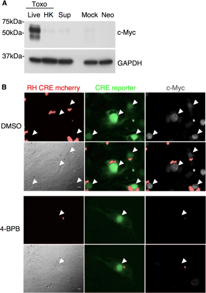
FIG 3.

Active invasion is necessary but not sufficient for c-Myc induction. (A) From left to right, HFFs were infected at an MOI of 1 with untreated (Live) or heat-killed (HK) Toxoplasma RH parasites (Toxo), treated with supernatant from Toxoplasma-infected culture (Sup), mock infected (Mock), or infected with Neospora (Neo). Cell lysates were prepared at 20 hpi and subjected to Western blot analysis of c-Myc and GAPDH levels as described for Fig. 1A. (B) Toxoplasma RH Cre mCherry parasites were treated with DMSO (upper set of 6 panels) or 4-bromophenacyl bromide (4-BPB; lower set of 6 panels) prior to infection. Cre reporter cells, which turn from red to green upon Cre-mediated recombination (Koshy et al. 21), were infected at an MOI of 1. c-Myc levels were determined by IFA 20 hpi. White arrows indicate nuclei of infected cells. All six panels in each group show the same field, with filters to detect mCherry (red; upper left), GFP (green; upper middle), c-Myc (white; upper right), phase (lower left), mCherry and GFP (lower middle), or mCherry and c-Myc (lower right). Scale bars in phase (lower left) panels indicate the distance of 10 μm.
