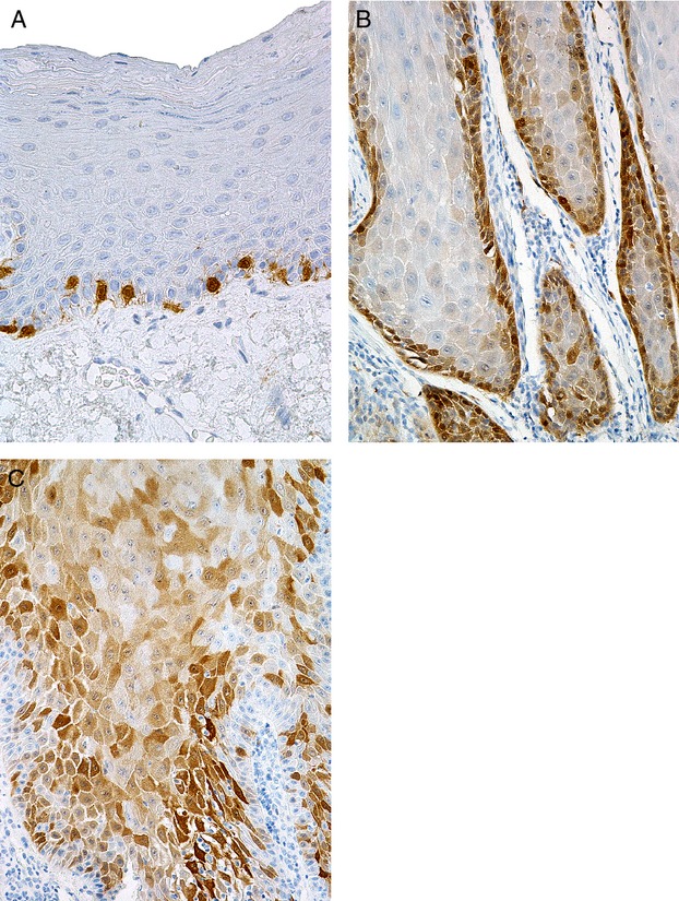
Figure 1.

Immunohistochemical expression of p16. (A) Normal epithelium: staining in occasional basal cells. (B) Verrucous carcinoma: staining in basal/parabasal cells. (C) Verrucous carcinoma: more extensive, mosaic staining in basal, parabasal and spinous cells.
