Figure 1.

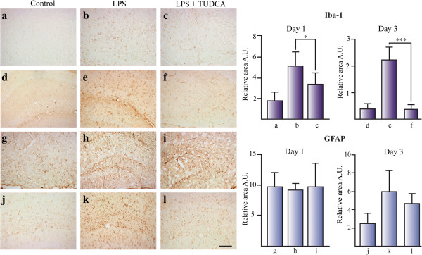
TUDCA reduces microglial activation in the hippocampus of LPS treated mice. The effect of TUDCA on glial activation was determined by the immunoreactive area for Iba-1 (for microglial cells) (a–f) and GFAP (for astrocytes) (g–l) related to total area in mice hippocampus icv injected with LPS. Section treatments are as follows: 1 day control (a, g), 1 day icv LPS (b, h), 1 day icv LPS + ip TUDCA (c, i), 3 day control (d, j), 3 day icv LPS (e, k), and 3 day icv LPS + ip TUDCA (f, l).*P <0.05, ***P <0.001. Scale bar represents 100 μm. The results represent the mean ± SD of at least 5 sections of 6 animals per group.
