Figure 5.
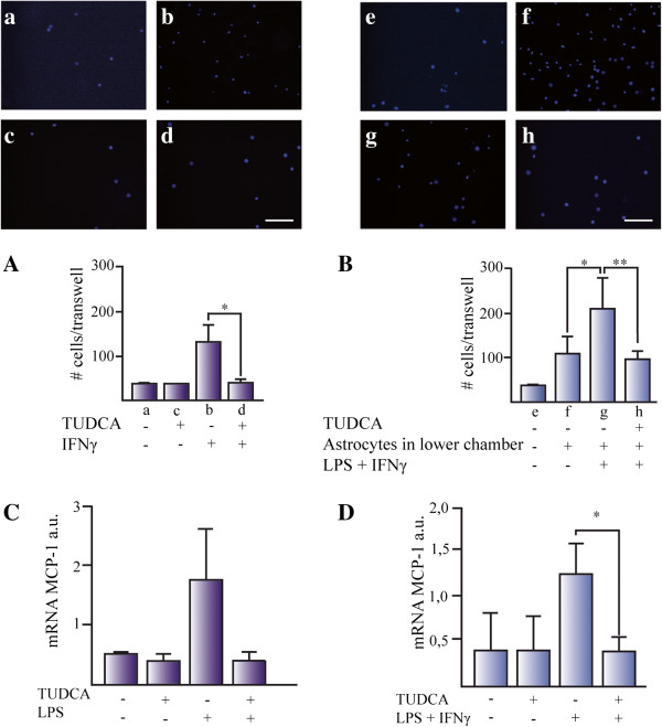
TUDCA reduces microglia cell migration in vitro. Microglial migration was studied using (A) IFN-γ and (B) conditioned media from proinflammatory stimuli-activated astrocytes after 24 h exposure. (A) IFN-γ was added in the lower part of the Transwell and microglial cells were seeded in the upper part. DAPI positive cells attached to the lower part of the Transwell were counted. Data represents the mean of the number of cells ± SD of at least five fields of three Transwells for each experimental group. Representative pictures were taken from cells treated with (a) control media, (b) control plus TUDCA, (c) IFN-γ, and (d) IFN-γ + TUDCA. (B) Microglial migration was studied in media conditioned for 24 h from: (e) control media without astrocytes, (f) untreated astrocytes, (g) astrocytes treated with LPS plus IFN-γ, and (h) astrocytes pretreated with TUDCA and treated with LPS plus IFN-γ. The expression of the mRNA for MCP-1 was determined by qPCR in (C) microglial cells and (D) astrocytes. The expression of mRNA for 36B4 and β-actin were used as loading control for microglial cells and astrocytes, respectively. The results represent the mean of the ratio between the expression of mRNA for iNOS/expression of mRNA for β-actin or 36B4 ± SD of at least three experiments in triplicates. *P <0.05. Scale bars represent 50 μm.

