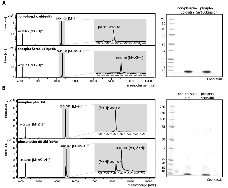
Figure 6. Quality control of phosphorylated Ubl (Parkin residues 1–76) and phosphorylated ubiquitin.
(A) MALDI–TOF spectra of non-phospho-ubiquitin (top panel) and phospho-Ser65 ubiquitin (bottom panel) after incubation with MBP–PINK1 after separation on a Mono Q column. (B) MALDI–TOF spectra of non-phosphorylated Ubl (Parkin residues 1–76) (top panel) and mixed Ubl species (~60% phosphorylated and ~40% non-phosphorylated) (bottom panel). (A and B) In total, 2 μg of the non-phospho-ubiquitin and phospho-ubiquitin (A) or non-phospho-Ubl and phospho-Ser65 Ubl (B) were resolved by SDS/PAGE, followed by staining with Colloidal Coomassie Blue for quality control.

