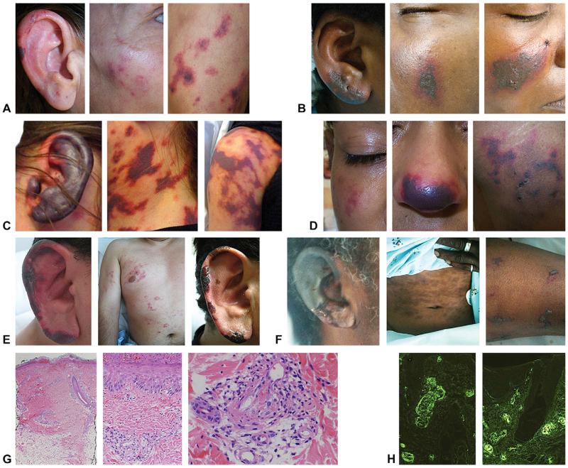
Fig 1.
A, Ear, cheek, and thigh of 46-year-old woman. B, Ear, left cheek, and right cheek of 57-year-old woman. C, Ear, neck, and thigh of 46-year-old woman. D, Cheek, nose, and thigh of 22-year-old woman. E, Ear (left) and trunk (middle) of 37-year-old man on admission, and same ear resolving 1 week later (right). F, Ear, abdomen, and arm of 50-year-old man. G, Representative histopathology from ear of patient (E) showing leukocytoclastic vasculitis at 32.5 (left), ×20 (middle), and ×40 (right) magnification. H, Representative direct immuno-fluorescence of ear biopsy specimen demonstrating positive IgM (left) and positive fibrin (right) in vascular and perivascular spaces.

