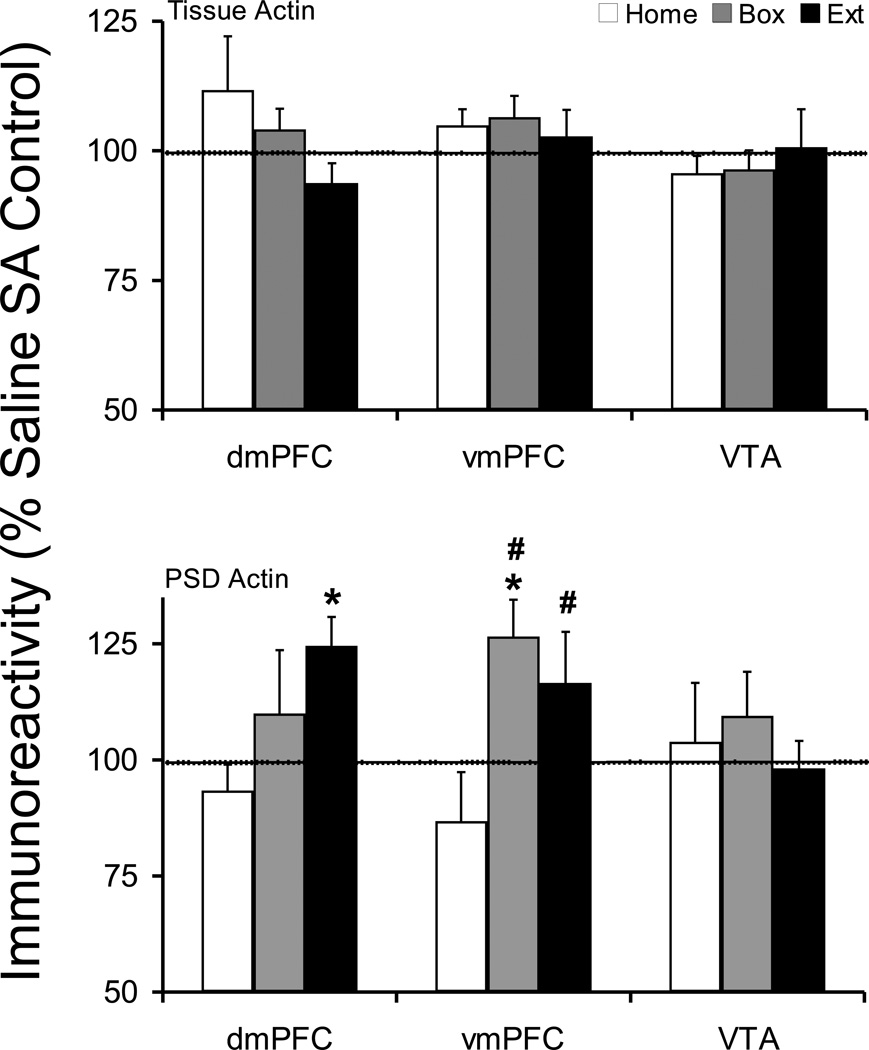
Figure 5.
Tissue and postsynaptic density (synaptosomal membrane fraction) levels of Actin protein after cocaine self-administration and Home, Box, and Extinction treatments. See Figure 1 legend for details. The tissue Actin protein was not changed following any post-SA treatment. However, dmPFC Extinction and vmPFC Box groups showed a significant increase in PSD Actin protein. Comparing cocaine self-administration groups across post-SA conditions, Actin levels were significantly higher in vmPFC Box and Extinction groups compared to Home group. *p<0.05 compared to respective saline control. #p<0.05 compared to Home cocaine group.

