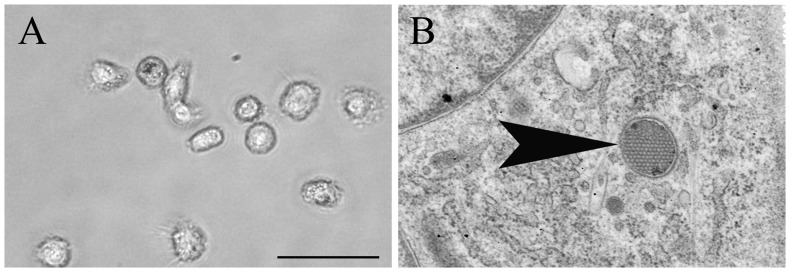
Figure 1. Morphological characterization of canine monocyte-derived dendritic cells at seven days in culture.
A) Cultured cells showing a typical dendritic cell-like morphology with long cytoplasmic processes. Phase contrast microscopy, bar size = 20 µm. B) Periodical microstructure (wasp nest-like structure; arrowhead) in the cytoplasm representing a distinct feature of canine monocyte-derived dendritic cells. Transmission electron microscopy, magnification = 25.000×.

