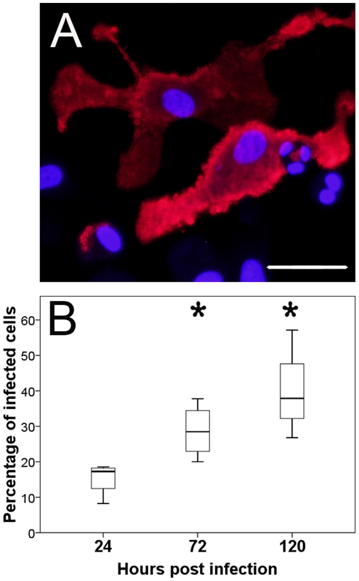
Figure 3. Detection of canine distemper virus (CDV) in monocyte-derived dendritic cells by immunofluorescence.
A) Infected monocyte-derived dendritic cells at 72 hours post infection (hpi) labeled with a CDV-specific antibody (red color). Nuclear staining with bisbenzimidine (blue color), bar size = 20 µm. B) Quantification of CDV-infected cells revealed a significant increase (*; p≤0.05) at 72 and 120 hpi. Box and whisker plots display median and quartiles with maximum and minimum values.

