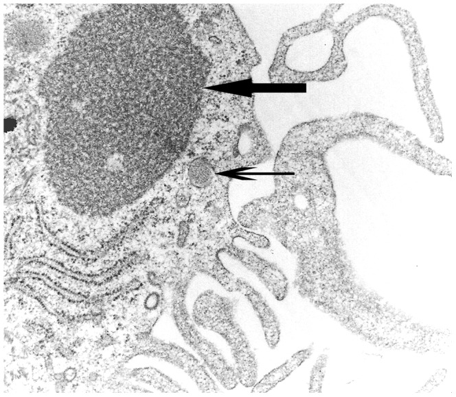
Figure 4. Ultrastructural analysis of canine distemper virus-infected monocyte-derived dendritic cells.

Accumulation of viral nucleocapsid in the cytoplasm (arrow). Note also periodical microstructure (arrowhead) and cytoplasmic processes representing features of dendritic cells. Transmission electron microscopy, magnification = 50.000×.
