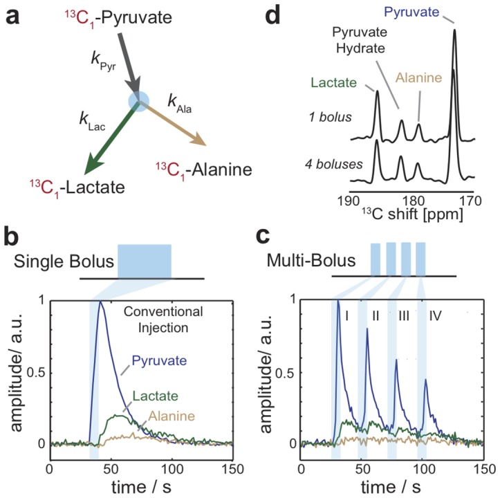
Figure 1. Multiplexing hyperpolarized magnetic resonance using multi-bolus tracer delivery.

(a) Metabolic scheme showing 13C label (red) transfer and respective rate constants for the dynamics of 13C-pyruvate, 13C-lactate, and 13C-alanine. (b) Conventional hyperpolarized magnetic resonance spectroscopy of 13C-pyruvate metabolism in mouse hind-limb skeletal muscle initiated by a single bolus event (I) with experiment repetition time of 1 s, and pulse flip angle of 15°. (c) Experimental demonstration of 4 bolus events (1–IV) with inter-bolus separation of ca. 30 s, experiment repetition time of 1 s, and pulse flip angle of 30°. (d) Summed 13C spectra from the conventional 1 bolus and multi-bolus measurements showing chemical shifts for observed hyperpolarized metabolites.
