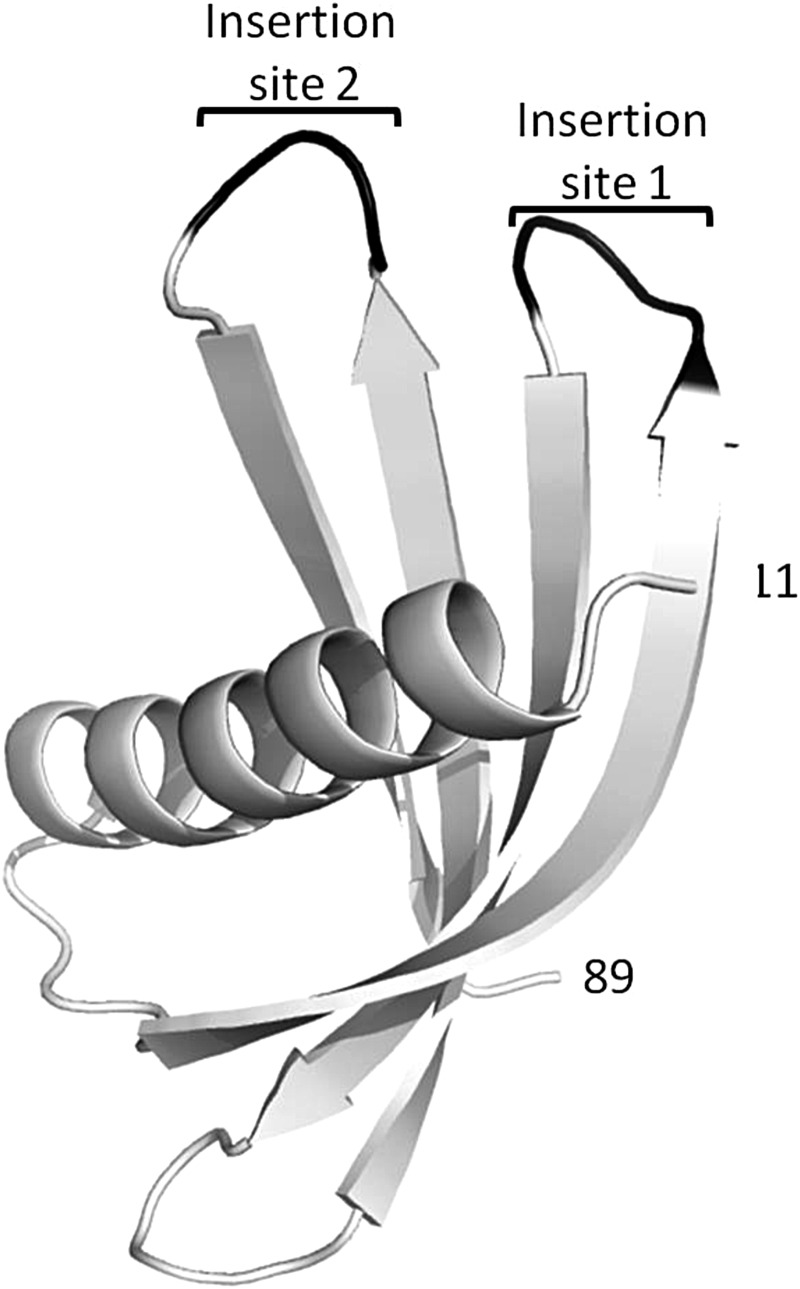
Fig. 3.
X-ray crystal structure of Adhiron92 scaffold (PDB ID no. 4N6T) at 1.75 Å resolution. The single alpha helix and the four anti-parallel β strands are shown in white with the insertion sites for library production shown in black. Residues 1–10 and 90–92 are not visible in the structure and are presumably disordered. The structure of Adhiron81 at 2.25 Å resolution (PDB ID no. 4N6U) is essentially identical.

