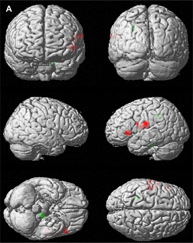
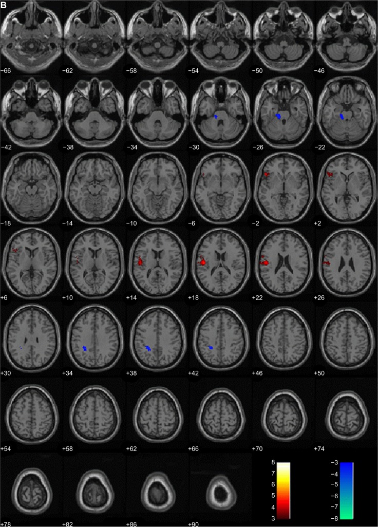
Figure 1.

Brain areas with changes in regional cerebral blood flow (rCBF) after 12 weekly sessions of group cognitive behavioral therapy in patients with panic disorder (n=14). (A) 3D-rendered images of rCBF changes. Red, increased rCBF; green, decreased rCBF. (B) Red, areas of increased rCBF, postcentral gyrus of left parietal lobe, precentral gyrus of left frontal lobe and inferior frontal gyrus of left frontal lobe; blue, areas of decreased rCBF, sub-gyral white matter of left limbic lobe and pons.

