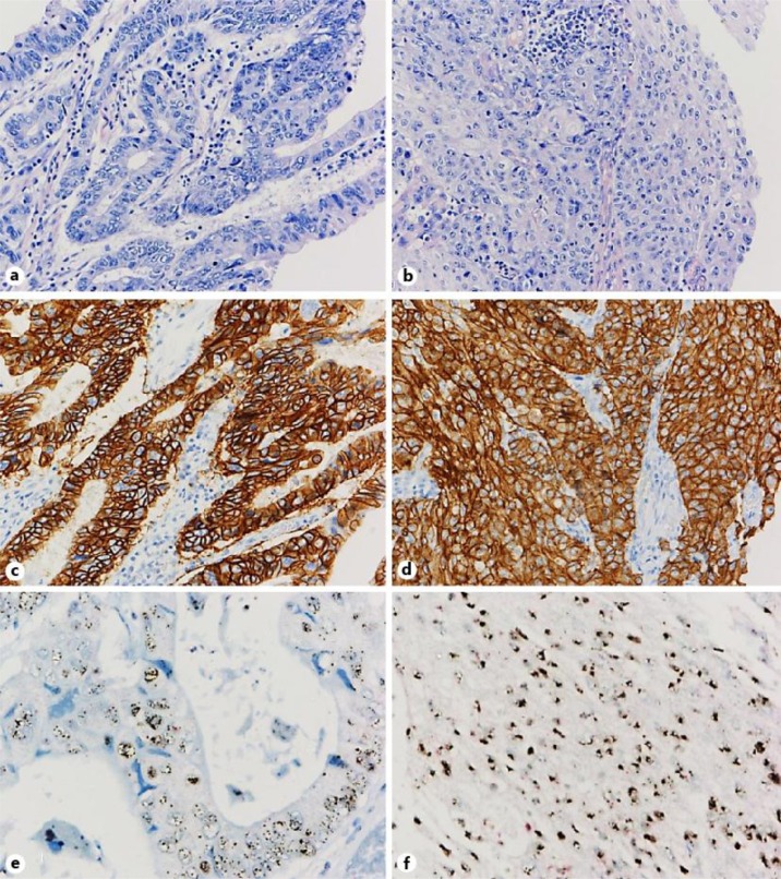
Fig. 2.
a, b The pathological diagnosis of the resected specimens was adenosquamous cell carcinoma with a mixture of both adenocarcinoma (a) and squamous cell carcinoma components (b). c, d Immunohistochemical analysis showed strong positivity for HER2 (HerceptestTM) in both components. e, f Dual-color chromogenic in situ hybridization revealed high amplification of the HER2 gene (INFORM HER2 Dual ISH; red, chromosome 17 centromere; black, HER2) in both components.

