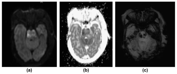
Figure 11.
A 24-day-old term neonate with confirmed bacterial meningitis. (a) Axial DWI image, (b) axial ADC map, and (c) axial mIP SWI image show DWI hyperintense signal abnormalities and restricted diffusion (low ADC values) affecting the pons representing ischaemic infarction as a complication of meningitis/vasculitis. mIP SWI image shows multiple hypointense foci within the pons representing haemorrhagic conversion/lesions within the infarcted region.

