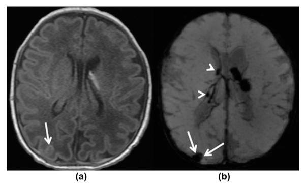
Figure 4.
A 6-day-old premature infant at 34 4/7 weeks of gestation with GMH grade I. (a) Axial, T1-weighted and (b) axial, mIP SWI images show a bilateral GMH, left greater than right. The small right GMH is only visible on the mIP SWI image (arrowheads on b). Additionally, small confluent haematomas are noted over the right occipital and posterior parietal convexities, most likely extra-axially located representing a subdural/subarachnoid haemorrhage (white arrows on a–b). They are easily visible on the mIP SWI image and could easily be missed on the T1-weighted images. The study was undertaken in an intubated child, hence the SWI signal intensity of the sulcal veins is increased due to the high oxygenation.

