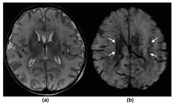
Figure 6.
A 5-day-old term newborn with severe hypoxic–ischaemic encephalopathy/injury. (a) Axial, T2-weighted and (b) mIP SWI images show moderate diffuse T2-hyperintense brain swelling, partially with a loss of the cortico-medullary differentiation in the occipital lobes and in the peri-rolandic region. mIP SWI image shows, in addition to findings seen on the T2-weighted image, prominent/dilated intramedullary and deep subependymal veins, which are typically seen in HIE/brain oedema. This “extra” information solidifies the diagnosis of HIE and may be helpful in predicting prognosis/outcome.

