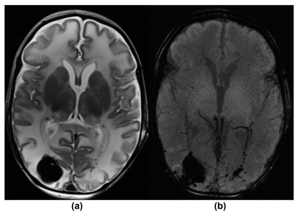
Figure 7.
A 12-day-old term newborn with hypoxic–ischaemic encephalopathy after hypothermia therapy. (a) Axial, T2-weighted and (b) mIP SWI images show mild global T2-hyperintense white matter brain oedema as well as a large haemorrhagic lesion in the right occipital lobe and additional small petechial haemorrhages in the left occipital lobe and subependymal region along the optic radiation, which are faintly seen on the T2-weighetd image. The sulcal veins are nearly completely “invisible” in this ventilated neonate. All haemorrhagic findings are seen in better detail on the SWI image compared to the T2-weighted image.

