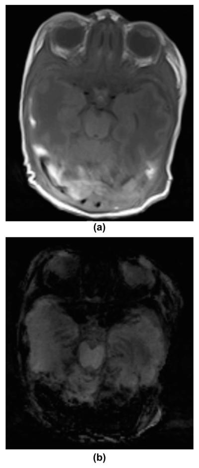
Figure 9.
An 11-day-old male infant with sinus vein thrombosis presented with perinatal depression, sepsis, and seizures. (a) T1-weighted MRI image demonstrates abnormal hyperintense signal within the transverse and sigmoid sinuses bilaterally, representing partial dural sinus thrombosis. (b) Axial mIP SWI image shows the typical matching blooming artefact of the intravascular venous thrombosis. No additional complications were noted.

