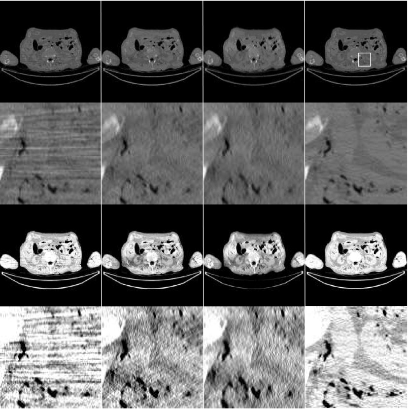
Figure 2.
Images reconstructed using low-dose CT clinical cadaver data. (First column) Conventional convolution backprojection reconstruction. (Second column) Proposed noise-weighted FBP reconstruction with spatial-domain filtering using ray-by-ray weighting. (Third column) Noise-weighted FBP reconstruction with spatial-domain filtering using view-by-view weighting. (Forth column) Gold-standard: conventional convolution backprojection reconstruction using regular-dose CT data. (First row) Display with a full gray-scale from minimum to maximum for each image. (Second row) Zoom-in of the first row; the region-of-interest is indicated in the upper-right image. (Third row) Display of the first row with a narrower gray-scale window. (Forth row) Display of the second row with a narrower gray-scale window.

