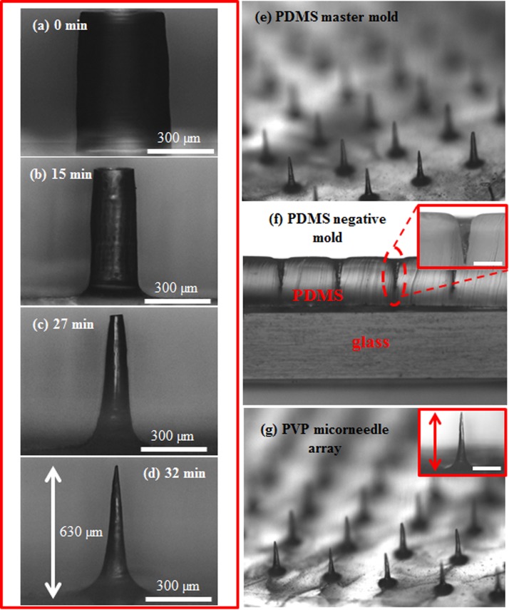
Figure 1.
(a) The initial micropillar with 400 μm in diameter and 700 μm in height. The optical images of PDMS micropillars at the etching time (b) 15, (c) 27, and (d) 32 min in the etching process without agitation. (e) The etched PDMS microneedle array. (f) The cross sectional view of the PDMS negative mold. (g) The PVP microneedle array. The scale bar in the insets of (f) and (g) is 300 μm.

