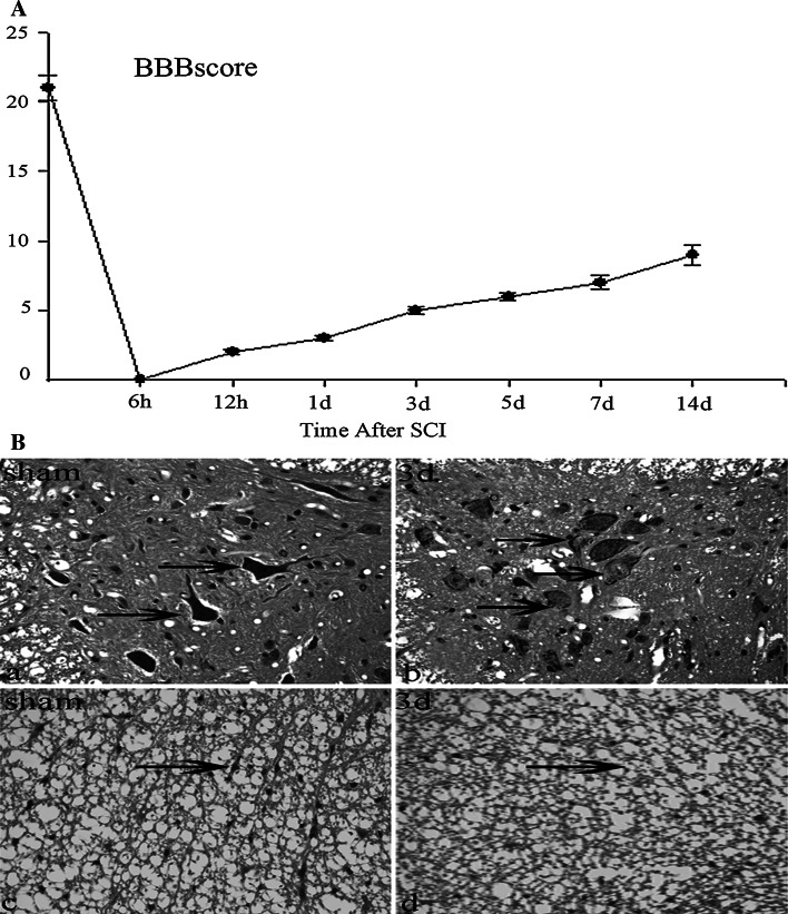
Fig. 1.
Behavior analysis and histology of the spinal cord of experimental animals. a Time course and degree of functional recovery in rats after SCI. Rats (n = 3) were killed at each time point (at 6 h and 12 h and at 1, 3, 5, 7, and 14 days post-injury). Data are reported as mean ± values of open-field locomotion BBB scores. b Histopathologic analysis of spinal cord from sham and injured rats at day 3 was used by H&E staining with paraffin histopathology sections. No lesion was seen in the sham group (a, c). Edema, vacuoles, irregularly shaped spaces, axon degradation, and disorders of the organization were evident in white matter at day 3 after injury (arrows in d). The majority of typical characteristics of neuronal apoptosis including nuclear fragmentation, nuclear disappearance, and nuclear pyknosis were found in gray matter (arrows in b). Scale bars, 20 μm (a–d)

