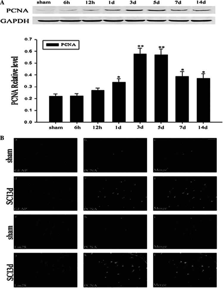
Fig. 6.
Association of LIN28 with the cell proliferation after SCI. Westernblot analysis of PCNA in spinal cord after SCI. The expression of PCNA was increased after SCI and peaked at day 3 (A). Double immunofluorescence staining is for PCNA, GFAP, and LIN28 in spinal cord after SCI (B). In adult spinal cord at 3 days after injury, sections labeled with PCNA (e) and GFAP (d) and the colocalization of PCNA with GFAP (yellow) were shown in the spinal cord (f). The majority of reactive astrocytes were PCNA-positive at 3 days after SCI (f). Moreover, there was colocalization between LIN28 and PCNA (l). However, we observed hardly any expression of PCNA in sham groups (b, h). Scale bars, 20 μm (b)

