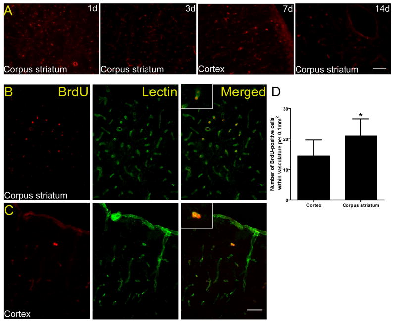
Figure 2.
In vivo migration of BMMNCs in rats that underwent 2VO. (A) Immunofluorescence staining for BrdU-labeled cells revealed that BMMNCs (red) migrated to both cortex (7 days) and white matter (corpus striatum area, 1, 3, and 14 days). (B) Double Immunofluorescence staining showed that BMMNCs (red) can be detected 2 weeks after transplantation in the area of corpus striatum. (C) The distribution of BMMNCs in the area of frontoparietal cortex. Most cells were incorporated into the vascular wall by day 14 after cell transplantation. Scale bars = 50 μm. n=3 at each time point. (D) Quantification showed that more vascular BrdU-positive cells were present in corpus striatum than in cortex. *p<0.05.

