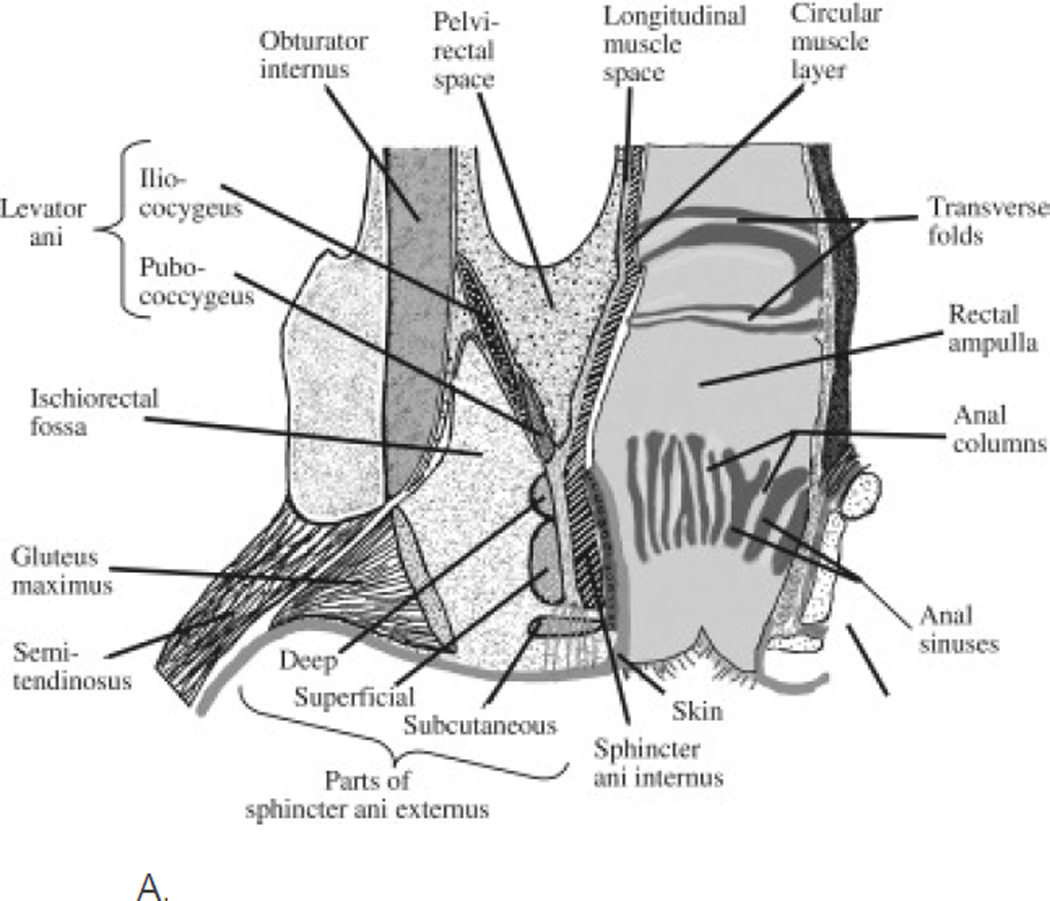
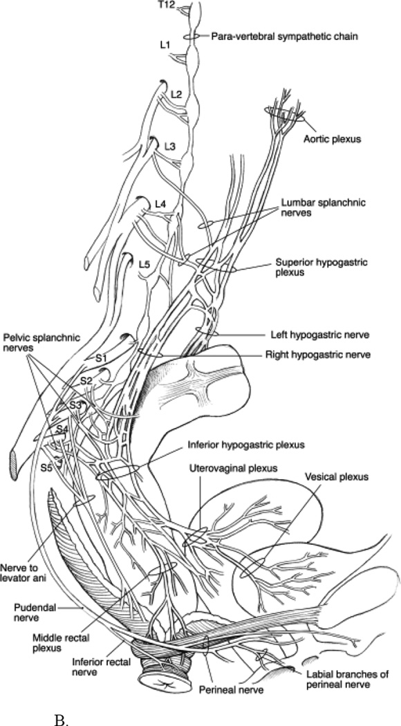
Figure 1.
A. Diagram of a coronal section of the rectum, anal canal, and adjacent structures. The pelvic barrier includes the anal sphincters and pelvic floor muscles. “Reprinted from Gastroenterology, 124, Bharucha, Fecal Incontinence, 1672-1685, 2003, with permission from Elsevier.”
B. Sympathetic, parasympathetic, and pudendal nerve supply to the anorectum. “Reprinted from Peripheral Neuropathy, 4th edition, Bharucha and Klingele, Autonomic and Somatic Systems to the Anorectum and Pelvic Floor, 279–298, 2005, with permission from Elsevier.”


