Figure 3.
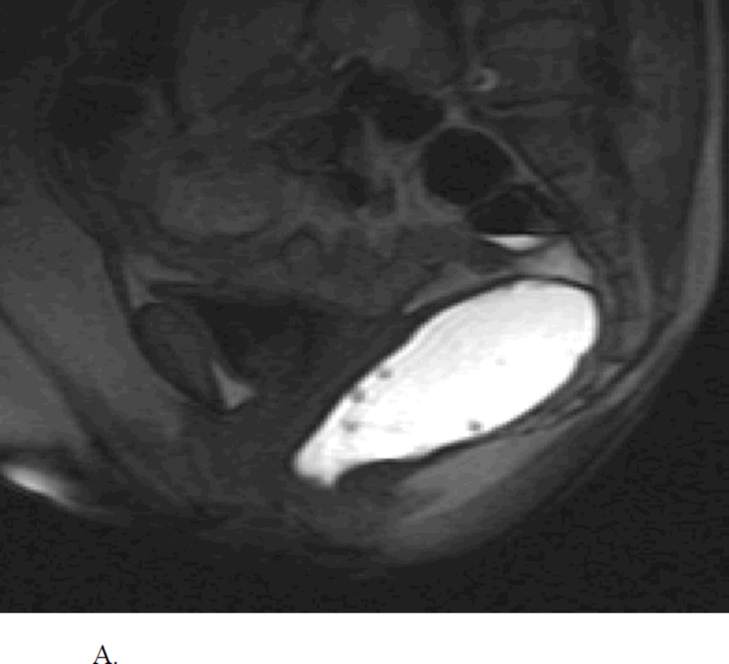
A. Normal MR Defecogram showing normal sized rectum filled with contrast and adjacent pelvic muscular and bony structures. Reproduced, with permission, from Roos J, Weishaput D, Wildermuth S, Willmann J, Marincek B, Hilfiker P, Experience of 4 Years with Open MR Defecography: Pictorial Review of Anorectal Anatomy and Disease, Radiographics, 2002, 22, 819.
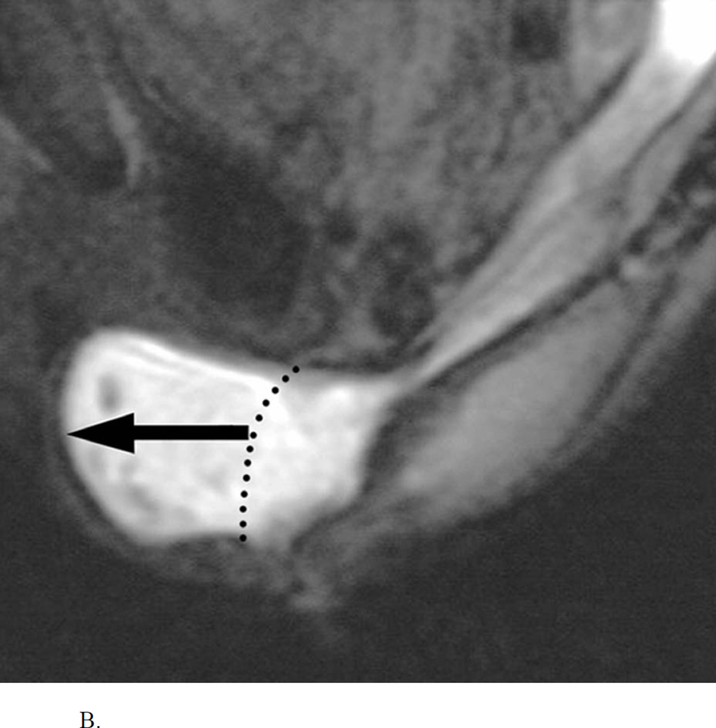
B. Abnormal MR Defecogram during an attempted defecation showing a large anterior rectocele (>4 cm). Reproduced, with permission, from Roos J, Weishaput D, Wildermuth S, Willmann J, Marincek B, Hilfiker P, Experience of 4 Years with Open MR Defecography: Pictorial Review of Anorectal Anatomy and Disease, Radiographics, 2002, 22, 825.


