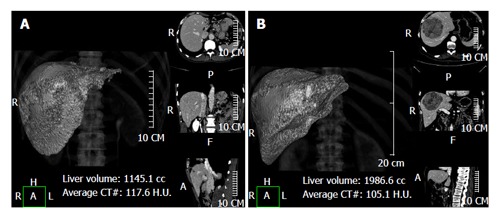
Figure 1.

Using a semi-automatic method liver analysis application provides a 3D and a multi-planar reconstruction of the liver. A case of hilar cholangiocarcinoma involving the left hepatic duct, with marked hypotrophy of the left lobe (type IIIb according to the Bismuth-Corlette classification) (A) and a case of hepatocarcinoma in segments 4-5-8 (B) are shown.
