Fig. 5.
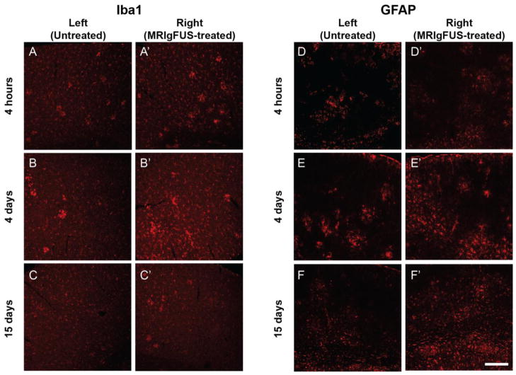
A time-dependent increase in Iba1 and GFAP staining in TgCRND8 mice treated with MRIgFUS. At 4 hours post-MRIgFUS treatment, Iba1 staining appears to be slightly increased in the MRIgFUS-treated cortex (A′) compared to the untreated cortex (A). Iba1-immunostaining intensity appeared to be elevated at 4 days (B, B′) and dampened by 15 days (C, C′). D, D′, GFAP expression did not seem affected by the MRIgFUS treatment at 4 hours. In contrast, at 4 (E, E′) and 15 (F, F′) days post-treatment, GFAP-positive staining was found to be increased on the right, MRIgFUS-targeted side compared to the left, untreated side. Scale bar: A–F′ = 200 μm.

