Fig. 8.
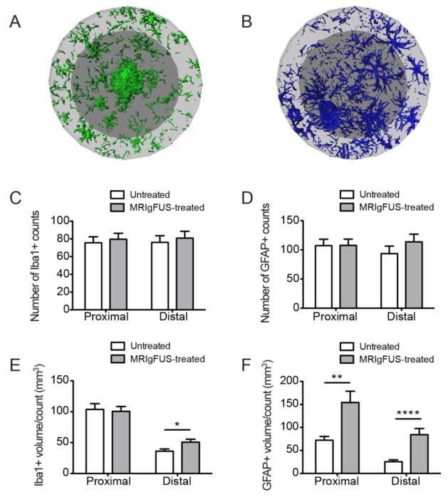
MRIgFUS treatment does not change the number of glial cells but increases their volume per cell. Representative images of microglia (A) and astrocytes (B) found in the proximal (dark grey) and distal (light grey) ROIs surrounding a single plaque in the MRIgFUS-treated cortex of a TgCRND8 mouse are shown. The number of Iba1- and GFAP-positive counts were not different between MRIgFUS-treated and untreated cortex (C, D respectively). E, The volume per Iba1-positive cell count, however, was increased significantly in the distal ROI of plaques in the MRIgFUS-targeted cortex compared to the untreated cortex. F, GFAP-positive volume per cell was significantly increased both in proximal and distal ROIs within the cortex treated with MRIgFUS. Mean ± SEM shown, *p<0.05, **p<0.01, ****p<0.0001.

