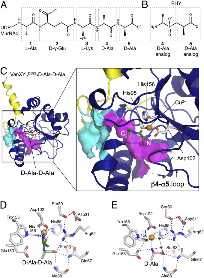
Fig. 2.
Ligand binding site. (A) Chemical structure of pentapeptide[d-Ala], with amino acids numbered from N to C terminus. (B) Chemical structure of PHY, phosphinate d-Ala-d-Ala analog, numbered according to the pentapeptide[d-Ala] it mimics. (C) VanXYCD59S•d-Ala-d-Ala complex. Solvent-exposed surface representation is shown in cyan and purple for putative ligand entry cavity. The N and C termini of d-Ala-d-Ala are indicated in white. (D) Interactions between VanXYCD59S, Zn2+, and d-Ala-d-Ala. (E) Interactions between VanXYCD59S, Cu2+, and d-Ala. Black spheres, water molecules.

