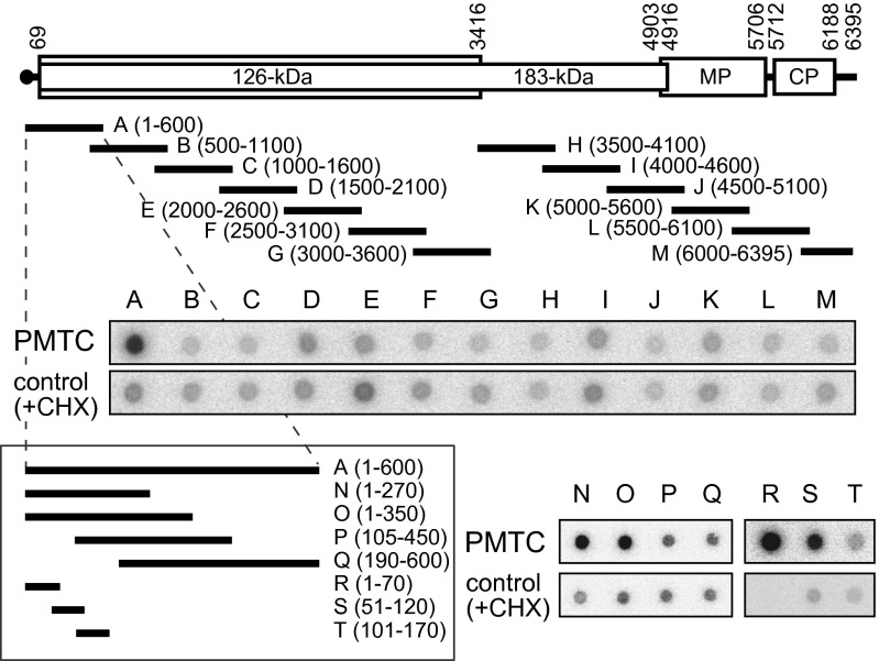
Fig. 2.
Mapping of the MNase-resistant region of TMV RNA in the PMTC. Schematic diagram of the TMV genome and the positions of cDNA fragments A–T. PCR-amplified double-stranded cDNA fragments A–Q and synthetic single-stranded DNA fragments R–T complementary to TMV RNA were blotted onto membranes and hybridized with RNA fragments recovered after MNase digestion of the PMTC fraction and 5′-32P-labeled. In the “control” panels, 32P-labeled probe RNA was prepared in the same way except that the reaction in mdBYL was performed in the presence of CHX, and was used for hybridization to the DNA fragments.

