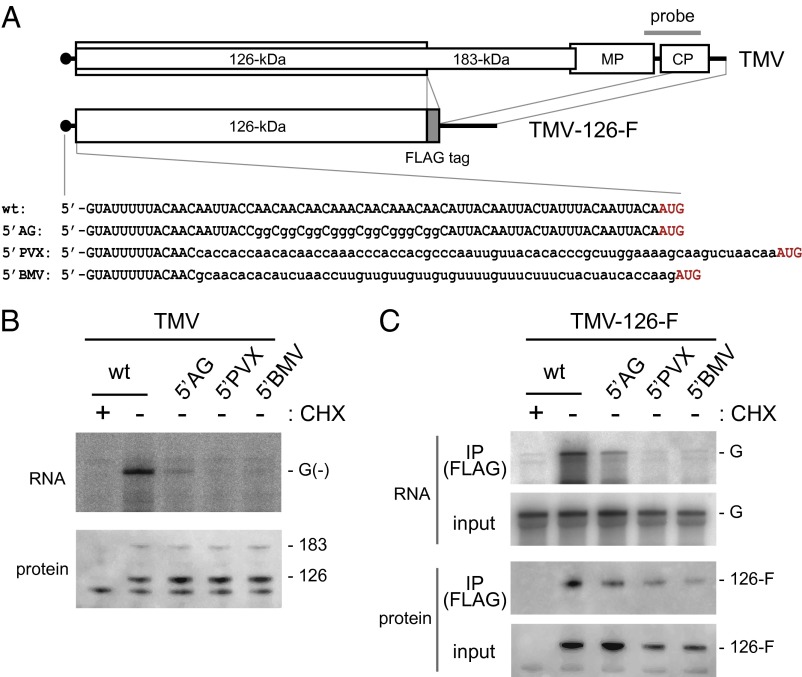
Fig. 4.
Effects of mutation of the 5′ UTR of TMV RNA on RNA replication and PMTC formation. (A) Schematic diagram of TMV RNA derivatives. The position of the probe (nucleotides 5,500–6,100) used for RNase protection and Northern hybridization is shown above the TMV genome. Nucleotide sequences of the 5′ UTRs of the TMV derivatives are shown in Lower. The initiation codon for the 126-kDa protein is shown in red. (B) In vitro translation and replication. Wild-type TMV and 5′ UTR-modified TMV RNAs were subjected to translation and replication reactions using BYL (CHX +, mock translation control). RNA was purified from the reaction mixtures and analyzed by RNase protection to detect negative-strand RNA (Upper). Production of the replication proteins was examined by Western blotting using anti-ToMV replication protein antibodies (Lower). Positions for protected RNA [G(–)] and the 126-kDa and 183-kDa proteins are indicated on the right. (C) Binding of the 126-kDa protein to genomic RNA. TMV-126F (wild type) and its 5′ UTR-modified RNA derivatives were translated in mdBYL (CHX +, mock translation control) followed by immunoprecipitation with anti-FLAG antibody-conjugated beads and elution with 3× FLAG peptide [IP (FLAG)]. RNA and protein were prepared from the input material and immunopurified fractions and detected by Northern hybridization using a 32P-labeled RNA probe complementary to the genomic RNA (Upper), as well as Western blotting using anti-126-kDa protein antibody (Lower). The positions of genomic RNA (G) and the FLAG-tagged 126-kDa protein are shown on the right.

