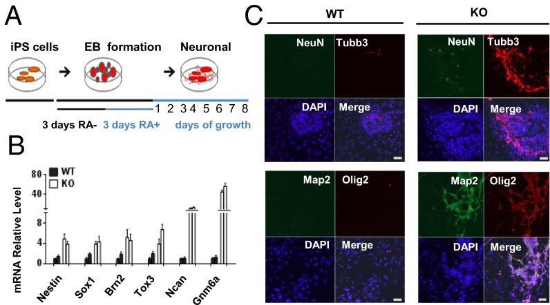
Fig. 3.
Increased expression of neuronal markers in KO iPS cells. (A) Protocol for neuronal differentiation. (B) mRNA expression analysis by qPCR of neuronal progenitor marker genes after 2 d of RA treatment in two KO iPS and two WT iPS clones. (C) Increased neuronal marker expression and arborization in KO iPS cells compared with WT iPS cells after 4 d of RA treatment. Nuclei were stained with DAPI. (Scale bar: 20 µm.)

