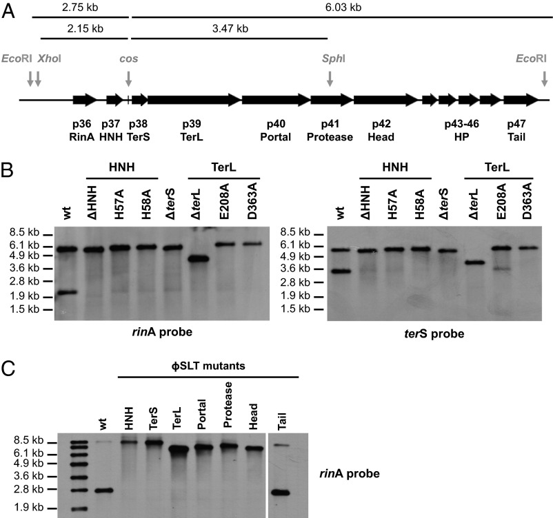
Fig. 4.
Role of the different ϕSLT mutants in phage packaging. (A) ϕSLT map. The relevant genes and the cos site are shown. (B and C) Lysogenic strains carrying the different phage mutations were exposed to MC, then incubated in broth at 32 °C. Samples were removed after 90 min and used to isolate DNA, which was digested with XhoI/SphI (B) or with EcoRI (C). DNAs were separated by agarose gel electrophoresis and transferred. Southern blot hybridization patterns of these samples were hybridized overnight with a rinA or terS (to the left and right of the cos site, respectively in A) phage-specific probes. In C, all samples were run on the same gel, but four lanes analysing mutants that are not included in the manuscript have been removed. Noncontiguous lanes are divided by a white line.

