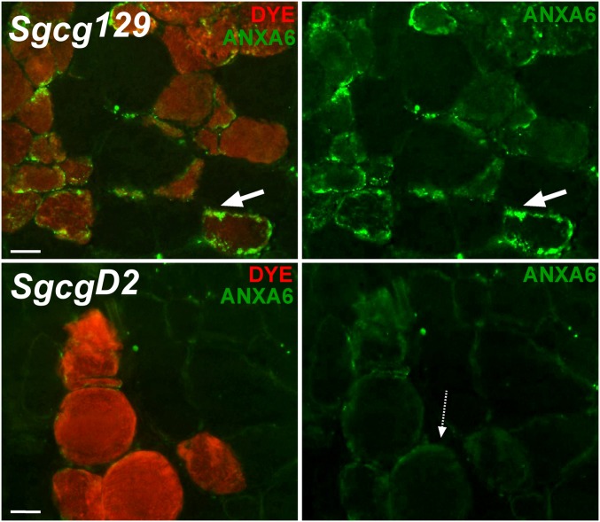
Fig. 5.
Altered annexin A6 intracellular localization in myofibers with disrupted membranes. Sgcg129 and SgcgD2 mice were injected with Evans blue dye (Dye, red) to mark damaged muscle fibers. Shown are representative images of Sgcg129 and SgcgD2 muscle immunostained with an antibody to the amino terminus of annexin A6 (green). Annexin A6 strongly localized in discrete patches on the periphery of dye-positive Sgcg129 fibers (top row, white arrow). Annexin A6 was only weakly present on the periphery of dye-positive SgcgD2 fibers (dotted arrow).

