Fig. 3.
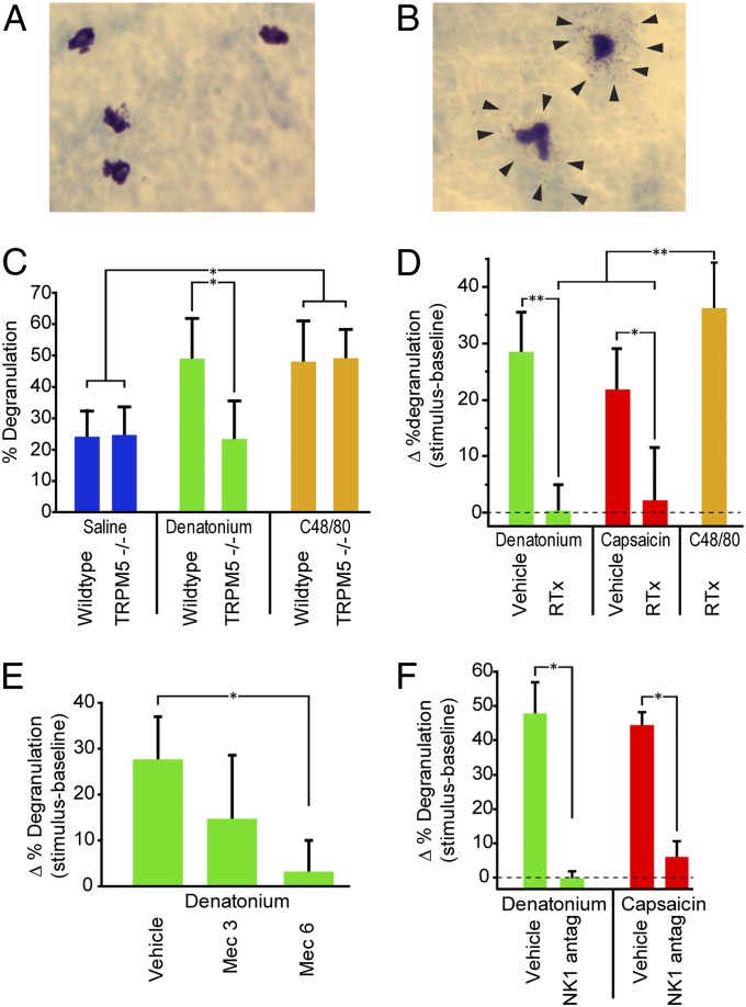
Stimulation of SCCs activates a proinflammatory pathway that triggers mast cell degranulation. Photos of (A) resting and (B) degranulated mast cells stained with acidified toluidine blue. Arrows point to granules forming a halo around the degranulated mast cells. (C) WT mice stimulated with 10 mM denatonium showed significantly more mast cell degranulation than TRPM5−/− animals (P < 0.05). Both WT and TRPM5−/− mice showed degranulation on exposure to the secretagague C48/80. (D) Mice treated with RTx to eliminate nerve fibers showed significantly less mast cell degranulation than vehicle-treated controls to both denatonium (P < 0.001) and capsaicin (P < 0.01) but were still able to respond to compound 48/80 (C48/80), which directly acts on mast cells to cause degranulation. (E) The nAChR antagonist mecamylamine (Mec) significantly reduced denatonium-induced mast cell degranulation at a dose of 6 mg/kg (P < 0.01). (F) The NK1 antagonist L732138 (5 mg/kg i.p.), which blocks responses to substance P, significantly reduces mast cell degranulation in response to both denatonium (P < 0.05) and capsaicin (P < 0.05). Bars represent mean + SEM. *P < 0.05; **P < 0.01 by one-way ANOVA with Tukey HSD test.

