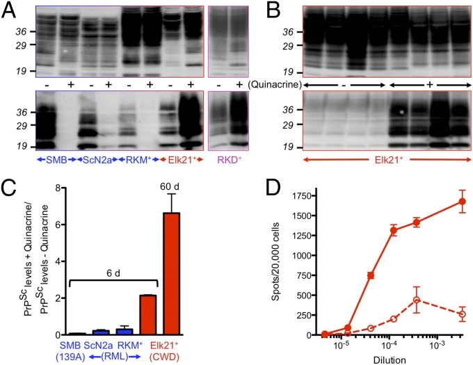
Fig. 1.
Discrepant effects of quinacrine on mouse and cervid PrPSc accumulation and prion replication. (A and B) Equal amounts of proteins from quinacrine treated (+) and untreated (−) cells were either not treated (Upper) or digested with PK to detect PrPSc (Lower). Blots were probed with mAb 6H4. (A) Cells were treated with 1 μM quinacrine for 6 d, except RKD+ were treated for eight passages. (B) Replicate plates of Elk21+ were treated with 1 μM quinacrine for 10 passages compared with untreated cells maintained for the same time. (C) Densitometric analysis of PrPSc levels in quinacrine-treated and untreated SMB (n = 3 pairs), ScN2a (n = 3 pairs), RKM+ (n = 3 pairs), and Elk21+ (n = 11 pairs) for 6 d, or Elk21+ for 60 d (n = 5 pairs). (D) Numbers of prion-infected Elk21+ cells following quinacrine treatment (solid line, filled circles) or no treatment (dashed line, open circles) determined by CPCA. Mouse, elk, or deer PrP indicated in blue, red, and magenta respectively.

