Fig. 2.
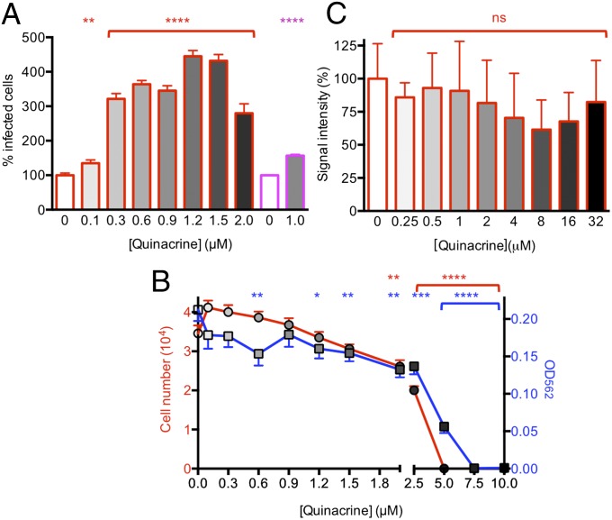
Effects of various quinacrine concentrations on CWD propagation and cell viability in Elk21+ and RKD+ cells, and during protein misfolding cyclic amplification. (A) Dose–response effects in Elk21+ (red) treated with quinacrine for 6 d, and RKD+ cells treated with 1 μM quinacrine for 60 d (magenta). (B) Effect of quinacrine on numbers of adherent Elk21+ (left y axis; red line, circles), and cell viability assessed by MTT assay (right y axis; blue line, squares). For each treatment, 16 replicates were assessed. The statistical difference of means was assessed by one-way ANOVA. (C) Densitometric analysis of western blotted PrPSc following PMCA performed in the absence or presence of quinacrine (n = 3 samples for each concentration). Error bars refer to standard errors of the mean (SEM). ns, P > 0.05; *P ≤ 0.05; **P ≤ 0.01; ***P ≤ 0.001; ****P ≤ 0.0001.

