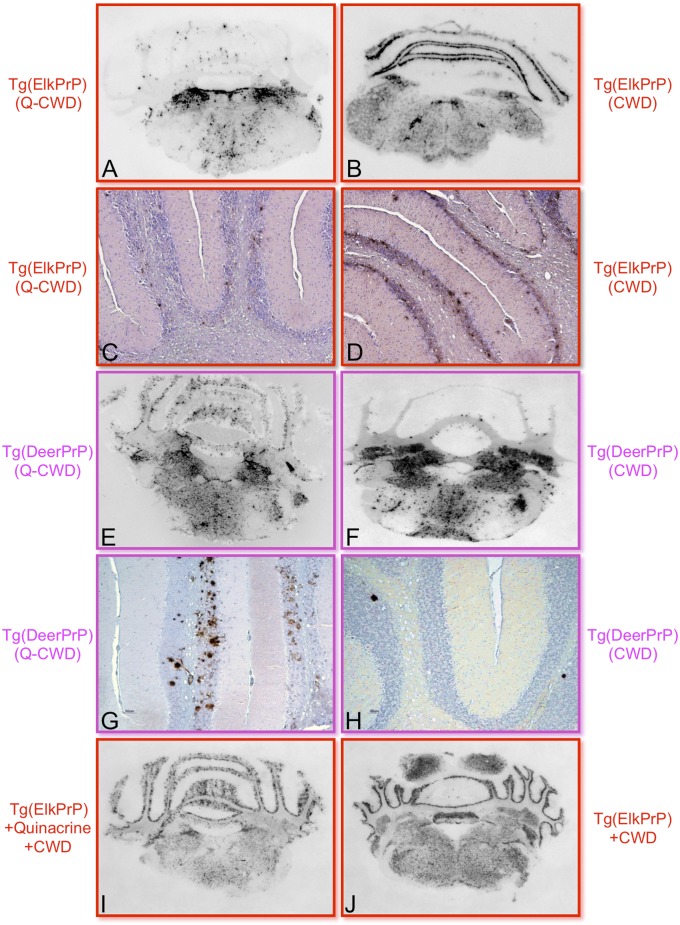
Fig. 5.
Q-CWD prions produce altered patterns of PrPSc deposition compared with untreated CWD prions. PrPSc deposition patterns in the region of the cerebellum assessed by histoblotting (A, B, E, F, I, and J) or immunohistochemistry (C, D, G, and H) in the brains of Tg(ElkPrP)5037+/− mice (A–D, I, and J) or Tg(DeerPrP)1536+/− mice (E–H) inoculated with Elk21+ treated with 1 μg/mL quinacrine (Left) or untreated Elk21+ (Right). (I and J) Histoblots of diseased, CWD-infected Tg(ElkPrP)5037+/− mice either treated (I) or not treated (J) with quinacrine.

