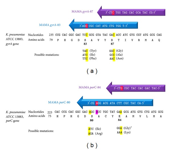
Figure 1.

MAMA-PCR primers for gyrA (a) and parC (b) mutation detection. Red highlighted nucleotides are the mismatched nucleotides at the 3′ end of each MAMA primer. Mismatches were positioned at the conserved nucleotides of each codon (highlighted by yellow) located at the 3rd nucleotide from the 3′ end of each primer, except for parC80 primer where the conserved nucleotide (1st nucleotide in the parC80 codon) was excluded from the MAMA primer and the alteration was situated at a nucleotide outside the coding region (pink highlighted nucleotide). Quality control strains with the expected mutations shown in the figure were used for the assay development and optimization except the mutation with ∗ which was not available.
