Figure 1.
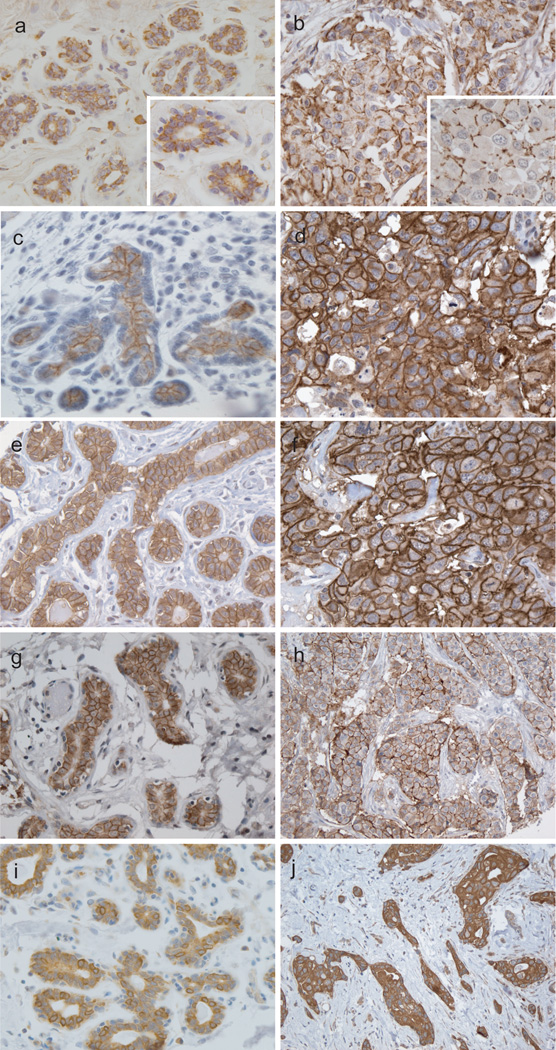
Expression of claudins in normal breast (A, C, E, G, and I) and invasive ductal carcinoma (B, D, F, H and J). Claudin 1 is expressed in the majority of luminal cells of terminal ductal lobular unit (A) and has scattered pattern in a basal-like carcinoma (B). Claudin 3 is expressed in the apicolateral side of the normal luminal epithelium (C) and the polarity is lost in a basal-like cancer (D). Predominantly apicolateral Claudin 4 expression in normal breast (E) and complete circumferential membranous staining in a basal-like carcinoma (F). Claudin 7 and 8 is also apicolateral in normal gland (G, I) and circumferential membranous in luminal subtype tumors (H, J).

