
Figure 3.
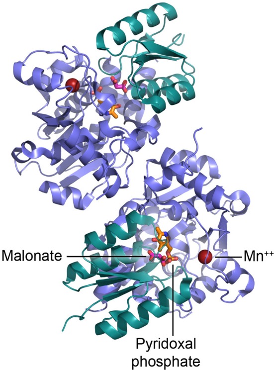
Dimeric structure of human SR and details of the active site. PLP is shown in orange, malonate in red, and Mn2+ as red sphere.
Official websites use .gov
A
.gov website belongs to an official
government organization in the United States.
Secure .gov websites use HTTPS
A lock (
) or https:// means you've safely
connected to the .gov website. Share sensitive
information only on official, secure websites.
Dimeric structure of human SR and details of the active site. PLP is shown in orange, malonate in red, and Mn2+ as red sphere.