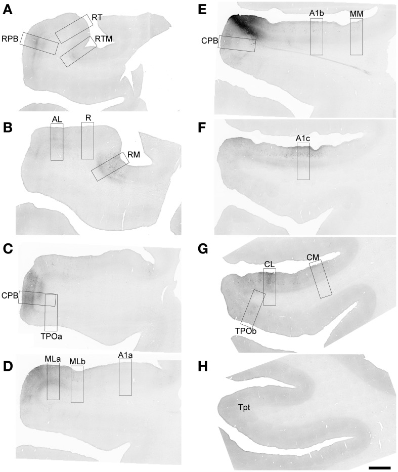
Figure 4.
FR labeling by the injection of ML in Case 2. Low magnification images of sections oriented from rostral (A) to caudal (H) along the STG. Black signal visible in each panel shows anterograde and retrograde transport from the FR injection in the caudal and lateral portions of ML (E). Rectangular selections correspond to higher magnification images in Figure 5. Scale, 2 mm.

