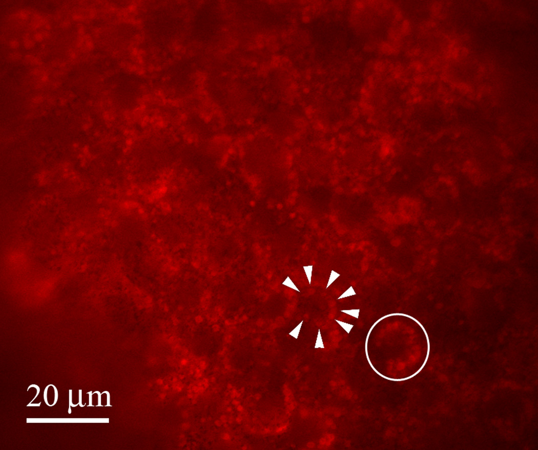
Figure 2.
Image of DiSBAC2(3) accumulation in cortical intracellular vesicles in the ectoderm of a Xenopus laevis embryo. Not all membranes are equal; thus a dye that accumulates in the plasma membrane of one cell type may accumulate in a different membrane of a different cell type. In this image, it is clear that the DiSBAC2(3) has accumulated in cortically located intracellular vesicles (arrowheads) of these ectodermal cells (single cell circled). The signal from these vesicles is very bright; thus it is not possible to use this dye for imaging the voltage of the plasma membrane of these cells. Scale bar = 20 μm.

