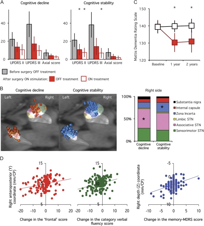
Figure 3. Postoperative parkinsonian disability and stimulating contact location in PD patients with vs without cognitive decline 1 year after surgery.
(A) Differences in parkinsonian activities of daily living (UPDRS part II), motor disability (UPDRS part III), and axial motor sign scores with bilateral subthalamic stimulation alone (OFF, solid red bars) or with drug (ON, empty red bars) in patients with PD who had postoperative cognitive decline (left panel) vs cognitive stability (right panel). The first bars (gray) represent the results obtained for the same scores in the off-drug condition before surgery in each group. (B) Location of the stimulating contacts of the patients with PD who had postoperative cognitive decline (left panel, red cylinders) vs cognitive stability (right panel, blue cylinders). The graph represents the relative proportion of stimulating contacts located within the different subthalamic subregions (sensorimotor, associative, and limbic) and surrounding areas (zona incerta/Forel field, internal capsule, substantia nigra pars reticulata). (C) Changes in the mean MDRS score in PD patients with (filled red squares) vs without (black squares) cognitive decline, 1 and 2 years after surgery. Note that in both groups cognitive status did not significantly change between the first and the second year postsurgery. (D) Relationship between the anteroposterior (Y) coordinate of the right stimulating contact and the change in the frontal score (left panel) and category verbal fluency (middle panel), and the depth (Z) of the right stimulating contact and the change in the MDRS score (right panel). Asterisks indicate significant differences between the 2 groups. CP = commissure posterior; MDRS = Mattis Dementia Rating Scale; PD = Parkinson disease; STN = subthalamic nucleus; UPDRS = Unified Parkinson’s Disease Rating Scale.

