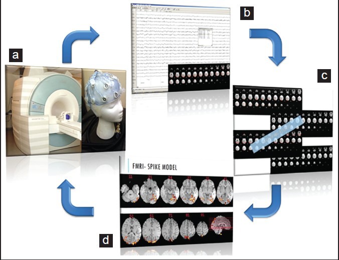
Figure 4.

Schematic showing analysis methodology in functional magnetic resonance imaging-electroencephalography (fMRI-EEG). EEG is acquired using a specialized system within the magnetic resonance imaging (MRI) machine while acquiring blood oxygenation level dependent (BOLD) sequences (a). Subsequently the EEG is analyzed for spikes and the corresponding BOLD fMRI change is detected (b). Multiple spike related BOLD signal changes are summated to improve the signal to noise ratio (c). The resultant summated signal is co-registered to the structural brain MRI to show the location of the summated BOLD signal change (d). This is the same patient in Figure 1. See how the summated signal in d corresponds to the lesion visualized with other structural and functional imaging modalities
