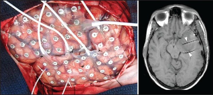
Figure 2.

An intraoperative photograph (left panel) showing left cerebral hemisphere covered by various subdural grid electrode arrays. Two depth electrodes are also seen (arrows) entering the parenchyma, which were inserted under neuronavigation guidance via lateral approach. A T1W axial magnetic resonance imaging (right panel) obtained following electrode implantation shows depth electrodes traversing the temporal lobe with tips located in the mesial temporal structures (arrow heads)
