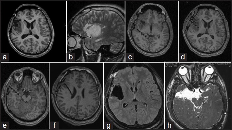
Figure 3.

The following example demonstrates the concept of epilepsy network. This 19-year-old boy presented with symptoms of “choking” sensation, followed by generalization (about 5-6 episodes a day since 5 years of age). He underwent a partial excision for a right insular low grade glioma at another hospital [Figures 1a and b]. This semiology changed 1 year ago to choking sensation followed by an aura of epigastric “rising,” followed by deviation of eyes toward left side. Video electroencephalography suggested an ictal onset in F8 and T4. Ictal single-photon emission computed tomography showed activity from the mesial right temporal lobe. Four depths were placed, one within tail hippocampus [c], one within the tumor [d], one within the hippocampus [e], and the 4th superior to the tumor [f]. Ictal onset was recorded from the electrode from hippocampus. Surgery included a complete excision of the insular glioma [g] and amygdalohippocampectomy with anterior temporal lobectomy [f]. Histopathology of the hippocampus showed evidence of mesial temporal sclerosis. Following surgery, patient had an Engel I outcome. Thus, even though the primary lesion was the insular low grade glioma, the hippocampal sclerosis was probably induced by the virtue of insular connections to the temporal lobe
