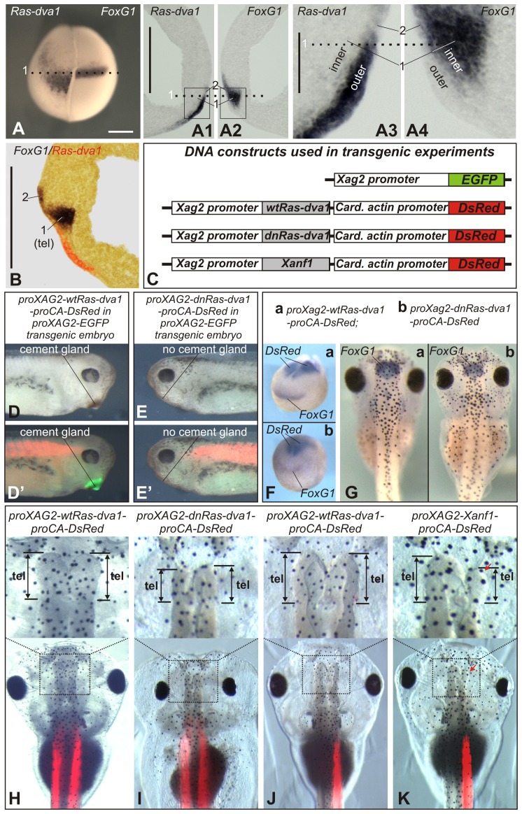
Fig. 1. Ras-dva1 is expressed in the non-neural anterior ectoderm and regulates forebrain development by a cell non-autonomous mechanism.
(A) In situ hybridization with dig-labeled probes to Ras-dva1 and FoxG1 on the left and right halves of the same embryo. Upon completing the in situ hybridization procedure, the two halves of the embryo were stacked together and photographed from the anterior with the dorsal side upward. The dotted line corresponds to the dotted lines in panels A1–A4. (A1,A2) Adjacent vibratome medial sagittal sections of the same embryo were hybridized separately with Ras-dva1 (left section) or FoxG1 (right section) probe. Anterior sides face each other, dorsal sides up. “1” indicates region of the inner layer in which only FoxG1 is expressed. “2” indicates region of the outer layer in which Ras-dva1 and FoxG1 are co-expressed. (A3,A4) Enlarged images of fragments squared in panels A1 and A2. (B) In situ hybridization with dig- and fluorescein-labeled probes to Ras-dva1 and FoxG1 on the vibratom medial sagittal section of the midneurula (stage 15) embryo. (C) Schemas of DNA constructs used to generate transgenic embryos shown in panels C–J. (D–E′) Whereas no cement gland inhibition is seen in control embryo of transgenic line bearing proXag2-EGFP construct and transfected by proXag2-wtRas-dva1-proCA-DsRed (D,D′), the embryo of the same line but transfected by proXAG2-dnRas-dva1-proCA-DsRed construct has no cement gland (E,E′). (F) No inhibition of FoxG1 expression is seen in the early neurula embryo bearing the control transgene (proXag2-wtRas-dva1-proCA-DsRed) (a). In contrast, a decrease of FoxG1 expression is observed in the embryo transfected with proXAG2-dnRas-dva1-proCA-DsRed (b). Whole-mount in situ hybridization with probes to both FoxG1 and DsRed. (G) Transgenic tadpole bearing the control proXag2-wtRas-dva1-proCA-DsRed construct has normal sized telencephalon marked by FoxG1 expression. At the same time, a reduction of the telencephalon and FoxG1 is seen in embryo bearing proXAG2-dnRas-dva1-proCA-DsRed construct. Transgenic tadpoles were selected by revealing DsRed fluorescence and hybridized in whole-mount with the probe to FoxG1. (H–K) The telencephalons (upper row) and whole heads (bottom row) of the 5-day tadpoles bearing transgenic constructs indicated on the top. Scale bars: 200 µm (A–A2,B), 40 µm (A3,A4).

