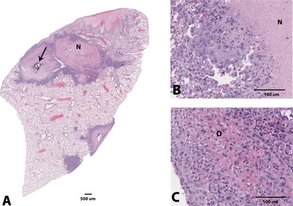
Figure 4.
Photomicrographs of the lung metastases in a mouse 12 weeks after undergoing surgical implantation of solid osteosarcoma tumor fragments. A: Variably-sized nodules composed of neoplastic osteoblasts surrounding a central region of coagulation necrosis (N) with occasional dystrophic mineralization (arrow), x0.5 magnification. B,C: Higher magnification of the coagulation necrosis (N); some neoplastic cells produce eosinophilic, osteoid-like matrix (O), x20 magnification. Hematoxylin and eosin staining.

